
An electrocardiogram, also called an ECG or EKG, records the electrical activity of the heart. In other words, it measures the electrical signals that the heart gives out, every time the heart muscles contract.
During an EKG, little patches called electrodes are placed onto your body. They detect the heart’s electrical signals, and send them into a machine which displays them as waves on a graph. The test is quick and painless, only taking a few minutes. Along with an echocardiogram, the EKG is one of the most valuable tools that a doctor can use to monitor heart health in Kawasaki Disease.
Doctors use EKGs to look at:
- Heart rate
- Heart rhythm
- Electrical activity
- Possible heart problems (Mayo clinic)
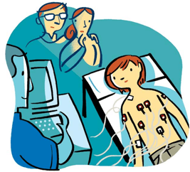
(Image Source: https://www.chp.edu/our-services/heart/patient-procedures/ekg)
Kawasaki Disease causes inflammation throughout the body as a result of an overreaction by the immune system. This inflammation often also affects the heart, changing the rhythm or electrical patterns that it gives out. So, doctors use electrocardiograms to track these changes and evaluate how Kawasaki Disease is affecting the heart. This is important because the inflammation can affect the heart itself, not just the coronary arteries. Therefore, using an EKG can help doctors detect the effects of inflammation on heart function.
However, unlike an echocardiogram, an EKG cannot directly show what is going on in the coronary arteries. So, often doctors use an EKG and echocardiogram together to monitor heart health, being able to track both the inflammation of the coronary arteries, and the overall function of the heart. (American Heart Association)


Leave a Reply